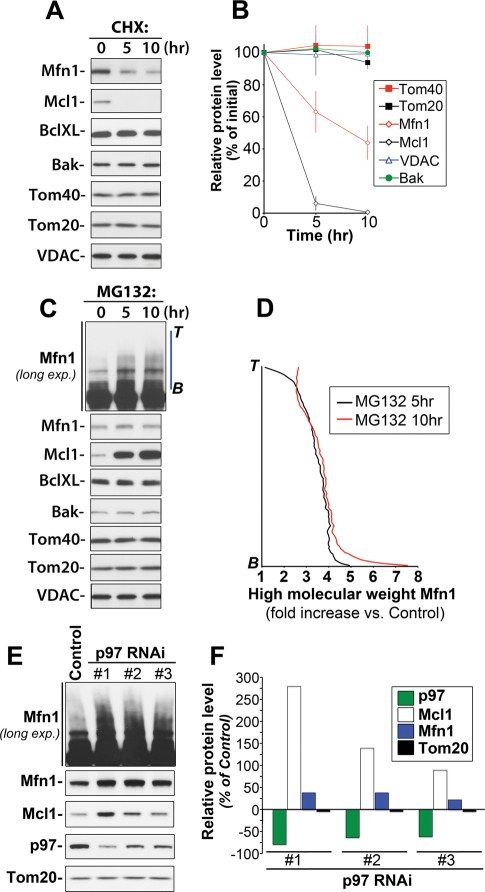
FIGURE 1:
Proteasome- and p97-dependent regulation of Mcl1 and Mfn1 stability. HeLa cells were treated with the protein synthesis inhibitor CHX (20 μg/ml) (A) or a proteasome inhibitor, MG132 (50 μM) (C), for 0, 5, or 10 h followed by Western blotting as indicated in the figure. In (B), relative protein levels in cells treated as described in (A) were quantified and plotted against the time of treatment with CHX [data represent the mean ± SD of three (VDAC, Bak, BclXL) or four (Mfn1, Mcl1, Tom20, Tom40) independent experiments]. In (D), the levels of high-molecular-weight species of Mfn1 were quantified in control cells and cells treated with MG132 for 5 or 10 h (along blue lines as shown in C; Mfn1-long exp.; B, bottom; T, top). The data shown represent fold increases of Mfn1 levels in different points of intensity plots versus the respective values in control samples. The data were normalized with control values at the respective points of intensity plots taken as 1. In panel E, total cell lysates obtained from cells transfected with 3 different p97 shRNAi constructs (#1, #2, and #3) or with a GFP shRNAi construct (Control) were analyzed by Western blot as indicated in the figure. In panel F, changes in the protein levels of p97, Mcl1, Mfn1, and Tom20 in p97 RNAi cells (clones #1, #2, and #3) were quantified and plotted as the percentage of the protein levels in control RNAi cells.

