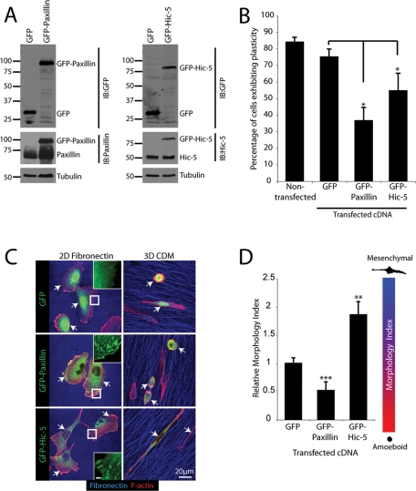
FIGURE 4:
Overexpression of paxillin and Hic-5 promotes MDA-MB-231 morphology transitions and inhibits plasticity. (A) Representative Western blot of MDA-MB-231 cells overexpressing pEGFPC1, pEGFPC1-paxillin, and pEGFPC1-Hic-5. (B) Quantitation of cells exhibiting plasticity during cell migration through 3D CDM for 16 h. (C) Representative immunofluorescence images, F-actin (red) and FN (blue), of cells spread on 2D and 3D ECM for 16 h in the presence of serum. Arrows indicate transfected cells. Inset images of the GFP channel indicate appropriate localization of pEGFPC1-paxillin and pEGFPC1-Hic-5 to adhesion contacts upon cell spreading on 2D FN. Inset scale bar = 2 μm. (D) Quantification of Morphology Index for cells migrating through 3D CDMs. Data represent mean ± SEM of a minimum of 20 cells from a minimum of four individual experiments. Statistical analyses of Morphology Index data was performed using a Kruskal–Wallis test followed by Dunn’s post hoc test, ** p < 0.001 and *** p < 0.0001.

