FIGURE 11:
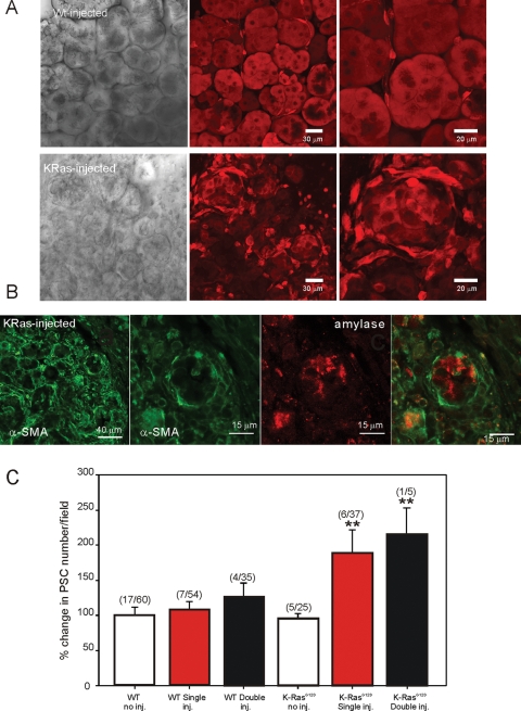
Morphology of lobules following cerulein injection. (A, top) Transmitted laser light image (left) from a lobule isolated from a WT animal injected with cerulein as detailed in Materials and Methods. Middle and right panels show MP excitation images of calcein fluorescence at 50× and 100× magnification, respectively, in WT lobules. (A, bottom) Transmitted laser light image (left) from a lobule isolated from a LSL-K-RasG12D animal treated with cerulein for 1 wk as detailed in Materials and Methods. Middle and right panels show MP excitation images of calcein fluorescence at 50× and 100× magnification, respectively, in LSL-K-RasG12D lobules. In contrast to WT animals, there is a severe disruption in acinar cell morphology together with an increase in periacinar cells. (B) Immunocytochemical staining of fixed lobules from LSL-K-RasG12D mice with antibodies against α-SMA and amylase to mark aPSC and acinar cells, respectively. Cells surrounding remnants of acinar cells are stained with α-SMA. (C) Analysis of the numbers of periacinar cells for the conditions stated. Numbers represent the number of animals/number of lobules at 50× magnification for each condition.

