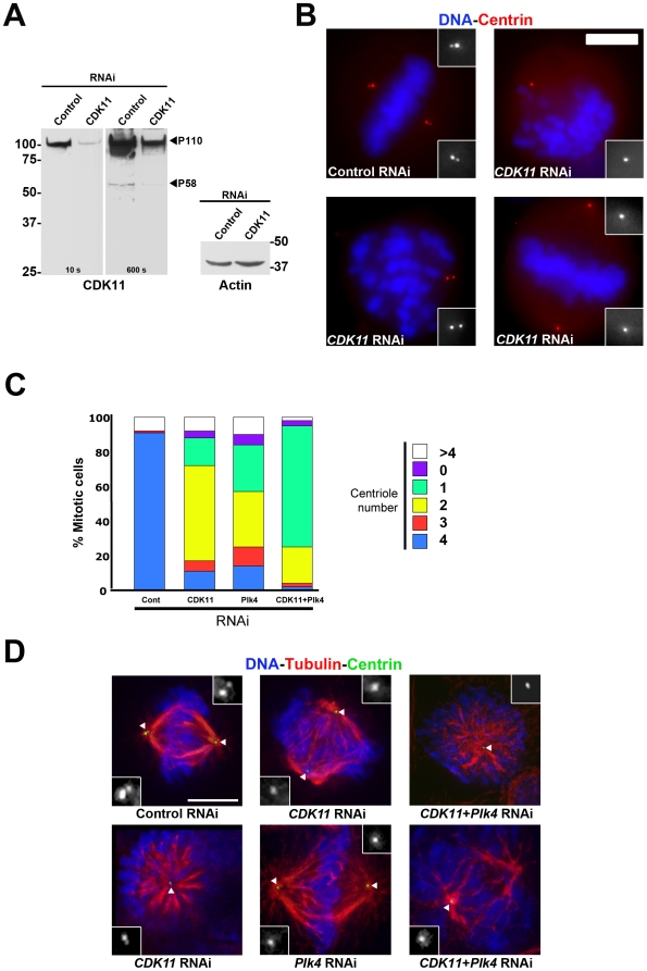
Figure 1. CDK11p58 depletion leads to a reduction in centriole number in mitotic HeLa cells.
Mitotic HeLa cells stably expressing a GFP-tagged centrin were subjected to control or CDK11 RNAi and analysed after 72 hours. A) Western blots showing CDK11p110 and CDK11p58 (with 10 and 600 seconds exposure times respectively) and actin protein levels are shown. The positions of CDK11p110 and CDK11p58 are indicated. B) Mitotic cells stained for DNA (blue) and centrin (red) following control or CDK11 RNAi. Some of the observed defects are shown here. The insets show a 3× magnification view of the centriole region in monochrome. The treatment is displayed at the bottom of each panel. Scale bar is 5µm. C) Quantitative analysis of the centriole distribution following control, CDK11, Plk4 or double RNAi in mitotic cells. See the reduced number of centrioles following CDK11 RNAi (also shown in Table S1). D) Example of the mitotic figures observed in control, CDK11, Plk4 and double (CDK11 and Plk4) siRNA–transfected cells. See also detailed analysed in Table S1. Centrioles are green, microtubules are red and chromosomes are blue. The insets show a 3× magnification view of the centriole region in monochrome (indicated by a white triangle in the merge panels). Bar is 5µm.

