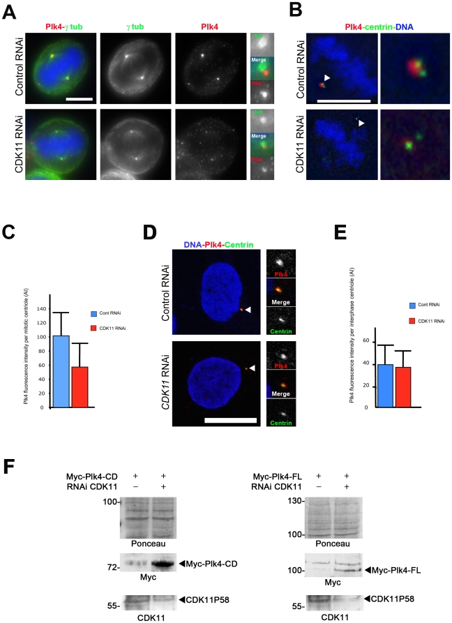
Figure 3. CDK11 depleted cells show diminished recruitment of the Plk4 protein at the mitotic centrosomes.
A) HeLa cells were transfected with control or CDK11 siRNAs and stained for Plk4 (red and right panels in monochrome) and γ tubulin (green and middle panels in monochrome). In the absence of CDK11p58, Plk4 signal is strongly reduced at the centrosome of mitotic cells. For each cell, one of the two centrosomal regions is enlarged on the left panel. Bar is 5µm. B) Single section of a control or CDK11 siRNA treated cell stained for centrin (green) and Plk4 (red). The insets show a 5× magnification of the spindle pole region that contains 2 centrioles. See the strong reduction of the Plk4 signal in the CDK11–depleted cell compared to the wild-type cell. Bar is 5µm. C) Graph showing the significant difference (p<0,0005) of Plk4 signal intensity (±SD) per mitotic centriole in control (blue) or CDK11 siRNA (red) transfected cells. D) Control (top) or CDK11 (bottom) siRNAs-transfected HeLa interphase cells stained for Plk4 (red), centrin (green). The right panels show a 3× magnification of the centrosomal region. Bar is 10µm. E) Graph showing the Plk4 signal (±SD) intensity per interphase centriole in control (blue) or CDK11 siRNA transfected cells (red). F) Analysis of exogenous Plk4 protein levels in CDK11 knock down cells. Myc-Plk4-CD (Catalytic Domain; amin-acids 1–638, left panels) and Myc-Plk4-FL (Full Length; amino-acids 1–970, right panels) expressing constructs were co-transfected with control or CDK11 siRNAs and the CDK11p58 (bottom panels) and Myc-Plk4 (middle panels) proteins levels were analysed by Western blotting 48 h following transfection. The membrane was stained by Ponceau S (top panels). In four different experiments, exogenous Plk4 proteins are hardly detectable after 48h hours in control cells whereas they are more stable following CDK11 RNAi.

