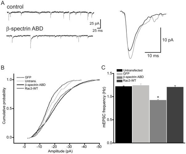
Figure 7. The ABD of βI-spectrin enhances single spine AMPAR-mediated synaptic responses.
A, mEPSCs from an untransfected control cell (upper trace) and a cell transfected with the ABD of βI-spectrin (lower trace) were recorded at −70 mV in the presence of TTX (1 µm). Average sized single mEPSCs recorded from the untransfected cell (black) and transfected cell (gray) are shown superimposed at right. B, Group data showing that neurons expressing the ABD of βI-spectrin had significantly greater mEPSC amplitudes, compared to untransfected controls or cells expressing GFP alone, suggesting an increase in AMPARs at these synapses (n = 7 neurons/group; p<0.05, K-S test). The increase in mEPSC amplitude produced by the spectrin ABD was mimicked in cells expressing CA Rac3, in which mEPSC amplitude was also larger than in controls or GFP expressing cells (p<0.05, K-S test). C, The frequency of mEPSCs in transfected neurons decreased significantly, suggesting fewer functional synapses (n = 7 neurons/group;* = p<0.05, t-test).

