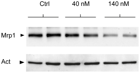
Figure 8. Western blot analysis of Mrp1 protein in the choroidal epithelium following bilirubin treatment.
Duplicate monolayers of choroidal epithelial cells were included for each experimental condition. Following daily basolateral treatment with 40 or 140 nM unbound bilirubin for 6 consecutive days, filters were treated in exactly the same conditions. The homogeneity in protein loading is reflected by the actin band revealed on the lower part of the membrane.

