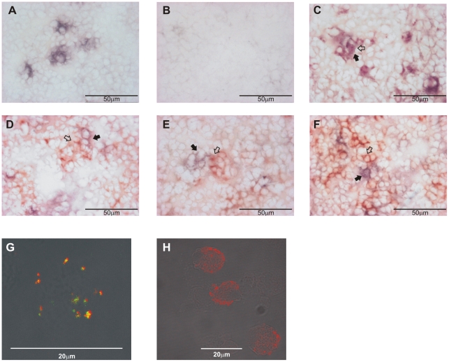
Figure 4. SITR protein is mainly expressed in myeloid cells.
A. Anti-SITR immunoreactivity (blue) in spleen of naïve carp. B. Anti-SITR immunoreactivity (blue) after pre-incubation of the anti-SITR antibody with the immunizing peptide (20µg/ml) in spleen of naïve carp. C. Double-staining for monocytes/macrophages (WCL-15; red) and SITR (blue) in spleen of naïve carp. D. Double-staining for neutrophilic granulocytes (TCL-BE8; red) and SITR (blue) in spleen of naïve carp. E. Double-staining for B cells (WCI-12; red) and SITR (blue) in spleen of naïve carp. F. Double-staining for thrombocytes (WCL-6; red) and SITR (blue) in spleen of naïve carp. G. Double-staining for monocytes/macrophages (WCL-15; green) and SITR (red) in macrophage-enriched cell fractions from head-kidney of naïve carp. H. Staining for SITR (red) in macrophage-enriched cell fractions from head-kidney of naïve carp. Typical red-stained (WCL-15+, TCL-BE8+, WCI-12+ or WCL-6+) cells are indicated with open arrows and typical blue-stained (SITR+) cells with closed arrows. Note that in panel C, it is difficult to distinguish between red- and blue-stained cells. Co-localization of both signals results in the indicated dark purple-stained cells.

