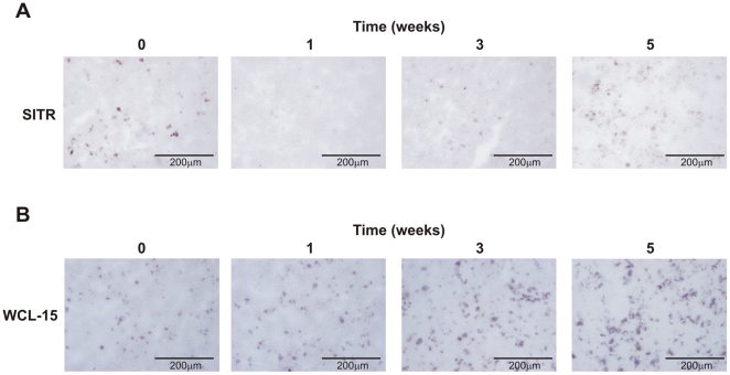
Figure 8. SITR protein expression during in vivo T. borreli infection.
A. Anti-SITR immunoreactivity (blue) in spleen tissue from non-infected fish (control) and at 1, 3 and 5 weeks post-infection with T. borreli parasites. B. Staining for monocytes/macrophages (WCL-15; red) in spleen tissue from non-infected fish (control) and at 1, 3 and 5 weeks post-infection with T. borreli parasites.

