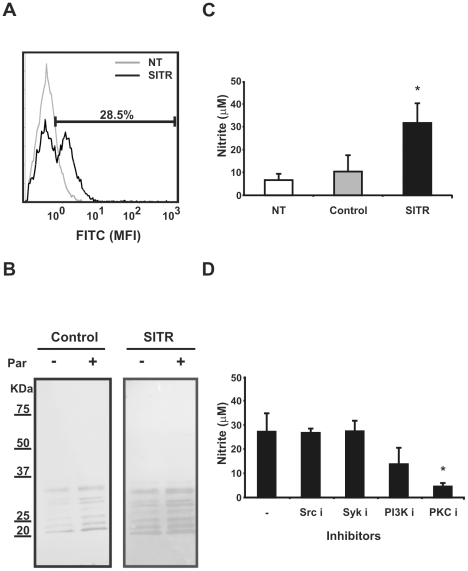
Figure 9. Overexpression of SITR in mouse RAW macrophages.
A. Intracellular SITR protein expression in RAW cells analysed by flow cytometry using anti-SITR antibody (1∶50). RAW cells were non-transfected (NT) or transfected with carp SITR. B. Western blot of cell lysates of TLR2ΔTIR- (control) and SITR-transfected RAW cells stimulated with live T. borreli parasites (Par, 0.5×10⁶ per well) for 15 min or left untreated as control. Tyrosine phosphorylation was evaluated using an anti-phospho tyrosine antibody. C. Nitrite concentration (averages ± SD of n = 5) in supernatants of non-transfected (NT), TLR2ÄTIR-(control) and SITR- transfected RAW cells determined by Griess reaction at 24 h. Symbol (*) indicates a significant (P≤0.05) difference compared with TLR2ÄTIR - transfected RAW cells. D. Nitrite concentration (averages ± SD of n = 3) in supernatants of SITR- transfected RAW cells pre-incubated for 30 min with inhibitors of Src kinase (Src i, PP2, 20µM), Syk kinase (Syk i, Piceatannol, 50µM), and PI3K kinase (PI3K i, LY294002, 50µM), PKC kinase (PKC i, Staurosporine, 1µM) or left untreated as control. TLR2ÄTIR-(control)- transfected RAW cells were used as negative controls (data not shown). Nitrite levels were determined by Griess reaction at 24h. Symbol (*) indicates a significant (P≤0.05) difference compared with unstimulated cells.

