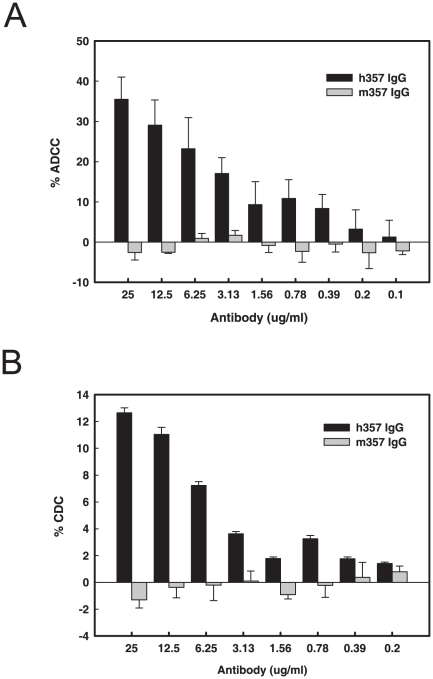
Figure 6. ADCC and CDC of h357 and m357 IgGs against transmembrane TNF-α-expressing cells.
Transmembrane TNF-α-expressing NS0 cells were incubated in the presence of different concentrations of h357 or m357 antibody for 1 hr. Subsequently, PBMCs for ADCC or human complement-rich serum for CDC were used as effector cells and the source of complement, respectively, and transmembrane TNF-α-expressing cells were used as targets. The cytotoxicity was calculated by measuring the amount of LDH released from the cytosol into the supernatant. Results are presented as percent cell lysis in antibody treated groups compared to 100% lysis in lysis buffer treated cell group.

