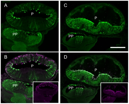
Figure 2. Localization of arrestins in the lamprey pineal organ.
A: Immunoreactivity to lamprey β-arrestin (green); B: merged image of immunoreactivity to β-arrestin (A) and parapinopsin (inset, magenta); C: immunoreactivity to lamprey visual arrestin (green) and D: merged image of immunoreactivity to visual arrestin (C) and rhodopsin (inset, magenta). β-arrestin is localized to the dorsal photoreceptor cells (A), whereas lamprey visual arrestin is localized to the ventral photoreceptor cells (C). P, pineal organ; PP, parapineal organ. Scale bar, 100 µm.

