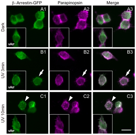
Figure 3. Light-induced translocation of β-arrestin with parapinopsin in cultured cells.
HEK 293S cells expressing lamprey parapinopsin and lamprey β-arrestin-GFP before irradiation (A) and 3 min (B) and 10 min (C) after irradiation with UV light. Panels Ai-Ci and panels Aii–Cii show fluorescence images of β-arrestin-GFP (green) and parapinopsin immunoreactivity (magenta), respectively. The inset of each figure shows the HEK 293S cells expressing lamprey visual arrestin-GFP (Ai–Ci inset, green) and lamprey parapinopsin (Aii–Cii inset, magenta) under the same light conditions. Panels Aiii–Ciii are merged images. Note that β-arrestin-GFP translocated to the cell membrane after UV irradiation (arrows) and subsequently appeared as granules also containing parapinopsin in the cytoplasm (arrowheads). In the HEK 293S cells expressing visual arrestin-GFP and parapinopsin, visual arrestin translocated to the cell membrane but was not observed in the intracellular granules after irradiation with UV light (inset). Scale bar, 10 µm.

