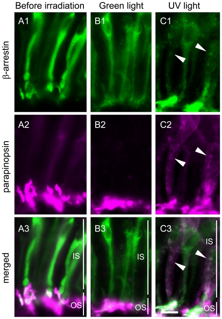
Figure 4. Light-regulated translocation of both β-arrestin and parapinopsin in the pineal photoreceptor cells.
Parapinopsin-containing photoreceptor cells in the dorsal region of the isolated pineal organs before irradiation (A), after irradiation with green light for 6 hours (B) and after irradiation with UV light for 6 hours (C). Panels Ai–Ci and panels Aii–Cii show β-arrestin (green) and parapinopsin (magenta), respectively. Panels Aiii–Ciii are merged images of both immunoreactivities. Many granules in the inner segment were stained by antibodies to both parapinopsin and β-arrestin (arrowheads in Ci–iii). IS, inner segment; OS, outer segment. Scale bars, 10 µm.

