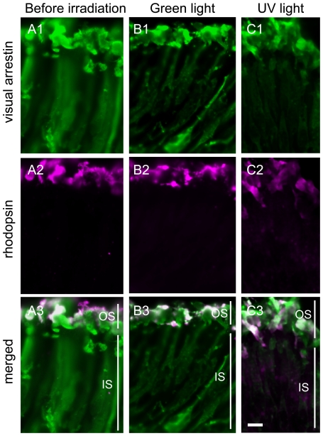
Figure 5. Light-regulated translocation of visual arrestin but not rhodopsin in the pineal photoreceptor cells.
Rhodopsin-containing photoreceptor cells in the ventral region of isolated pineal organs before irradiation (A), after irradiation with green light for 6 hours (B) and after irradiation with UV light for 6 hours (C). Panels Ai-Ci and panels Aii–Cii show visual arrestin (green) and rhodopsin (magenta) immunoreactivities, respectively. Panels Aiii–Ciii are merged images of both immunoreactivities. The results indicate that visual arrestin translocated to the outer segments, but no granules containing rhodopsin and visual arrestin were observed. IS, inner segment; OS, outer segment. Scale bars, 10 µm.

