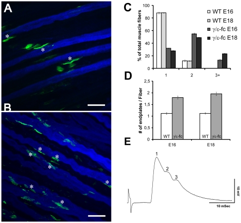
Figure 7. Presence of Multiple Endplates.
(A) Slow myosin (blue) and Alexa 488-labeled α-bungarotoxin (green) staining of E16 WT and (B) γ/ε-fc diaphragm. The asterisks mark the location of endplates on individual muscle fibers. The γ/ε-fc diaphragm presents multiple endplates on individual muscle fibers (scale bar: 25 µm). (C) Percentages of analyzed muscle fibers of WT and γ/ε-fc embryos displaying one, two, or three or more endplates at E16 and E18. (D) Average number of endplates per muscle fiber in E18 wild type (1,12±0,03, white) and γ/ε-fc (1,95±0,07, gray) embryos (n = 30 muscle fibers, 3 embryos per point, p<0,001). All data are represented as mean ± SEM. (E) Electrophysiological evidence of polyinnervations of an endplate in the diaphragm muscle of an of an E19 γ/ε-fc embryo. The endplate potential consists of 3 separate components, which had the same stimulus threshold for initiation.

