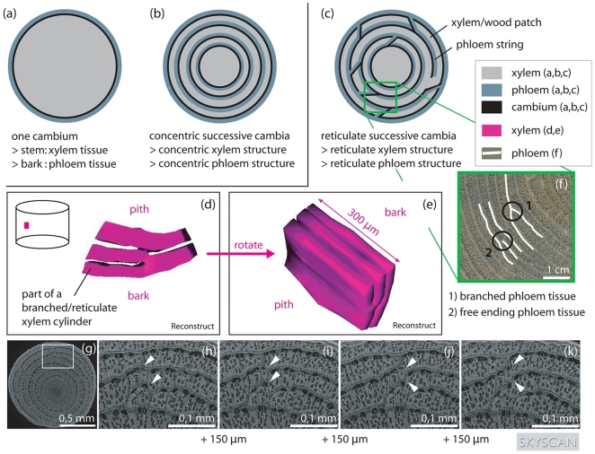
Figure 1. Spatial structure of the xylem and phloem tissue in Avicennia in comparison to other trees.
Schematic view of a stem disc from (a) a tree with only one cambium, (b) a tree with successive cambia organised in concentric cylinders, giving rise to a stem disc with concentric circles of xylem tissue, phloem tissue and cambium circles and (c) Avicennia, having a reticulate organisation of its cambia and transport structure. The spatial structure of Avicennia is depicted by smoothed surface images after three-dimensional reconstruction in Reconstruct [42] (d,e). Only a small part of the xylem tissue has been visualised. It can be seen that Avicennia had a complex network of xylem patches that fused at certain heights of the tree and joined different patches at other parts of the stem. The connections between xylem patches changed rapidly with vertical distance. This is shown by the white arrowheads in the four serial micro-CT-scan images of an outside zone of an Avicennia marina stem disc, produced by a SkyScan 1172 scanner (g-k). Each section is 150 µm apart from the previous. On the micro-CT-images (g-k) the wide light grey bands are xylem tissue while the dark small strings are phloem tissue. The complexity of the internal structure can be expressed by the sum of the locations of free ending and branched phloem tissue (f) per stem disc surface area.

