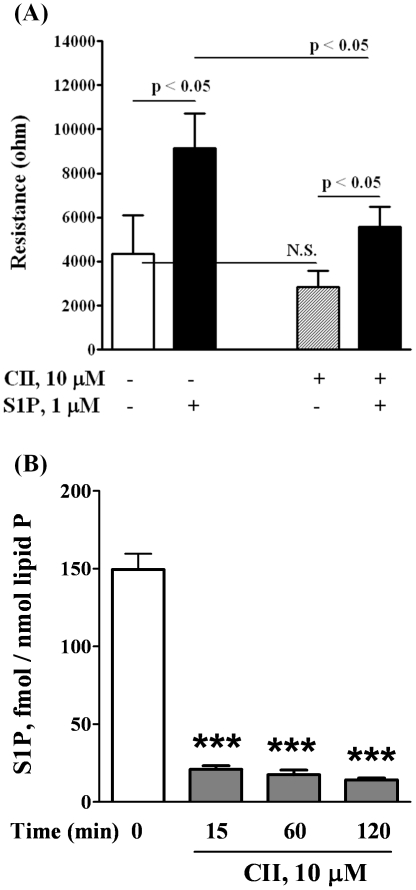
Figure 1. CII, inhibitor of SphK, decreases both the migration of HPAECs in a scratch assay in vitro and the content of S1P in HPAECs.
HPAECs grown to ∼95% confluence in 35 mm dishes were starved for 3 h in 0.1% FBS in EBM-2 without growth factors and treated with 10 µM of CII. Monolayers were scratched, and challenged with medium containing 0.1% BSA or 1.0 µM S1P complexed to 0.1% BSA. A shows the migration of cells into a “wound” that was scratched and exposed to S1P. B shows the decrease of S1P content in cells as measured by LC/MS/MS (see Methods) after lipid extraction from harvested cells. The values are mean ± S.E.M for three independent experiments each performed in triplicate (*** - p<0.001 in comparison to T = 0 min).

