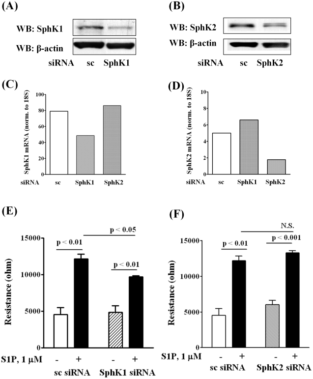
Figure 2. Silencing of SphK1, but not SphK2, decreases HPAEC migration by S1P in wound healing ECIS assay.
HPAECs (∼50% confluence) grown on 35-mm dishes or on gold electrodes (10–4 cm2) were transfected with either scrambled siRNA or SphK1 siRNA or SphK2 siRNA (25 nM, 72 h), then starved for 3 h in 0.1% FBS in EBM-2 without growth factors. Cell lysates (20 µg total protein) were subjected to SDS-PAGE on 10% precast Tris-Glycine gels and Western blotted with anti-SphK1 (A) and SphK2 (B) antibody as described under “Experimental procedures”. Western blot is representative of three independent experiments. Total RNA was isolated from control and SphK1 siRNA (C) and SphK2 (D) transfected cells, and Real-time PCR was performed in a Light Cycler using SYBR Green QuantiTect. Control and transfected cells were wounded on the gold electrodes (E and F) as described under “Experimental Procedures” prior to S1P (1.0 µM) challenge. Measurement of transendothelial electrical resistance (TER) using an electrical cell substrate impedance-sensing system (ECIS) for 16 h after wounding the cells on the gold electrode and exposure to 1.0 µM S1P was carried out. Values are the mean ± S.E.M. for three independent experiments in triplicate.

