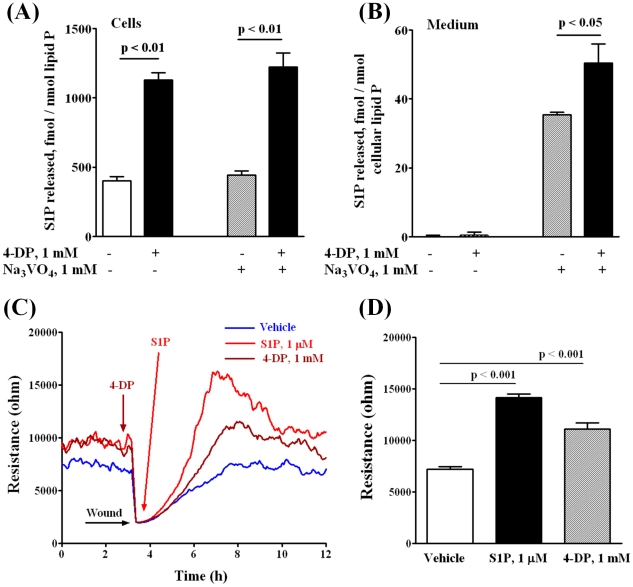
Figure 4. 4-Deoxypyridoxine increases intracellular content of S1P in HPAECs and stimulates cell migration in a wound healing ECIS assay.
HPAECs (∼90% confluence) grown on 35-mm dishes or on gold electrodes were starved for 3 h in 0.1% FBS in EBM-2 without growth factors and treated with 1 mM 4-DP for 6 h in the same medium. A, B – 4-DP increases intracellular content of S1P ( A ) and S1P release into the medium ( B ). Ortho-vanadate was added 30 min before lipid extraction in a fresh medium. S1P content in cells and medium was determined by LC-MS/MS as described under “Experimental procedures”. ( C, D ) - Control and 4-DP-treated (1 mM for 30 min) cells were wounded on the gold electrodes as described under “Experimental Procedures” prior to S1P (1 µM) challenge. D shows the changes in TER (ohms) in vehicle and 4-DP or S1P-treated cells at 4 h after wounding. Values are the mean ± S.E.M. for three independent experiments.

