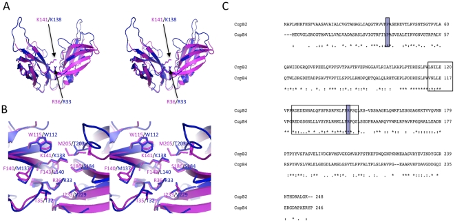
Figure 5. Structural comparison between CupB2 and CupB4 chaperones.
A) Homology model of CupB4 (blue) predicted based on E. coli PapD and P. aeruginosa CupB2 (magenta). B) Enlarged views of the putative active sites of CupB2 (magenta) and CupB4 (blue). Side-chains of the residues that are located 4 Å away from the invariant Arg and Lys are shown in stick representation. C) Sequence alignment between CupB2 and CupB4. Strictly conserved Arg, Lys and F1 strand - loop - G1 strand region, which are thought to be important for chaperone-subunit interaction, are highlighted. “*”, “:” and “.” indicate the strictly, moderately and weakly conserved residues, respectively.

