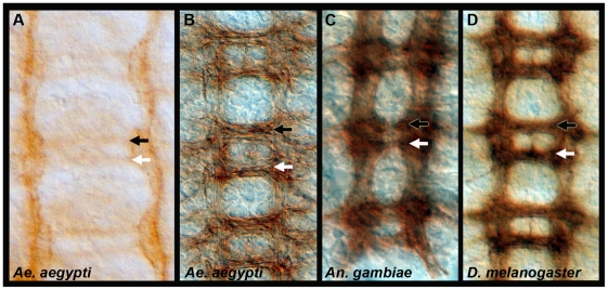
Figure 1. Development of the Ae. aegypti embryonic ventral nerve cord.
Anti-acetylated tubulin staining (A–C) marks the developing axon tracts in 52 hr. (A) and 56 hr. (B) Ae. aegypti embryos. By 56 hrs. (C), the Ae. aegypti nerve cord resembles that of a 33 hr. An. gambiae embryo and a St. 16 Dr. melanogaster nerve cord (BP102 staining is shown in D). These time points in the three respective species correspond to germ-band retracted embryos in which segmentation is obvious and organogenesis has initiated. Filleted nerve cords are oriented anterior up in all panels. The anterior commissure is marked by a black arrowhead, and a white arrowhead marks the posterior commissure.

