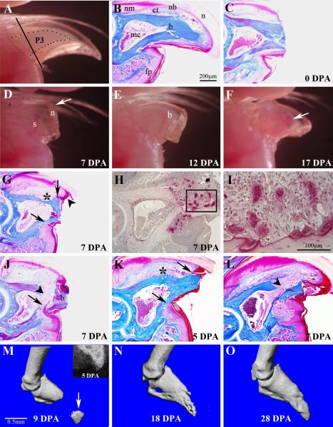
Figure 1. External morphology and wound healing during adult digit tip regeneration.
A) The distal region of the adult digit tip is comprised of the tip of the P3 skeletal element (outlined) that is visible through the opaque nail plate, the surrounding connective tissue and the nail plate. The plane of amputation is indicated by the solid line. B) Sagittal section through an unamputated digit tip shows a triangle shaped bone (b) that tapers to a pointed tip and with a bone marrow cavity (mc) localized in the proximal region. A thin band of connective tissue (ct) that is more prominent dorsally surrounds the P3 bone. Epidermal derivatives include the proximal multi-layered nail matrix (nm) that extends distally into the nail bed (nb) and underlying the nail plate (n). The digit fat pad (fp) is located ventrally in the proximal region of the digit tip. C) Sagittal section of the digit tip immediately after amputation. Amputation is through the distal cortical bone and nail bed, but leaves the marrow cavity intact. D) External view of a regenerating digit tip at 7 DPA showing distal elongation (arrow) of the nail plate (n), with no remarkable change of the stump bone (s). E) External view of a regenerating digit tip at 12 DPA showing outgrowth of a prominent digit blastema (b). F) External view of a regenerating digit tip at 17 DPA showing a regenerate that has the general shape of the terminal phalanx. Vasculature associated with ossification is visible through the nail plate (arrow). G) Histological section of a regenerating digit at 7 DPA showing an increase in the connective tissue dorsal to the bone, epidermal attachment to the dorsal and ventral bone surfaces (arrows), and distal overgrowth of the dorsal epidermis (arrowhead). Significant bone degradation (*) is observed at this stage. H) TRAP staining of 7 DPA regenerates identifies osteoclasts that are specifically localized to the amputated stump. I) Higher magnification of the region within the box in H showing some osteoclasts directly associated with bone and other osteoclasts that appear to be within the wound mesenchyme. J) Histological section of a regenerating digit at 7 DPA showing epidermal migration (arrow) through a region of eroded stump bone. This mode of wound closure causes a re-amputation and sloughing of the amputated bone (b). Bone erosion opens the distal region of the marrow cavity (arrowhead) allowing marrow-derived cells to enter the wound. K) Amputation at the very distal digit tip creates a very small amputation wound. At 5 DPA the epidermis remains attached to the dorsal and ventral bone surfaces (arrows) and there is evidence of bone erosion (*) at the level of the marrow cavity. L) In very distal amputations at 7 DPA, wound closure is still incomplete, the bone enclosing the marrow cavity is completely eroded and the marrow cavity is contiguous with the amputation wound (arrowhead). M-O) Longitudinal imaging of a regenerating digit tip. M) MicroCT scan at 9 DPA shows a re-amputated distal bone fragment (arrow) prior to being sloughed off. An X-ray image of the digit tip at 5 DPA (inset) shows the distal bony stump and an intact marrow cavity. N) MicroCT scan at 18 DPA shows the regenerated digit tip is nearly complete. O) MicroCT scan at 28 DPA showing the digit tip completely regenerated digit.

