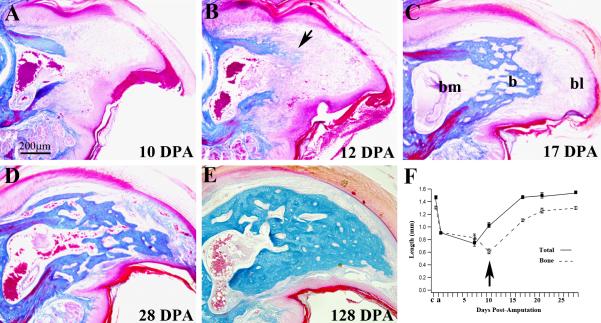
Figure 3. Re-differentiation.
Mid-sagittal histological sections of the regenerating digit tip stained with Malory's triple stain. A) 10 DPA blastema shows no histological evidence of differentiation. B) Direct ossification (arrow) was first observed in 1 of 6 samples analyzed at 12 DPA. C) At 17 DPA all sample had regenerated new trabecular bone (b) that capped the distal region of the bone marrow cavity (bm) proximal to the digit blastema (bl). D) At 28 DPA the distal region of P3 is regenerated with an interlacing network of new trabecular bone that is histologically distinct from the original cortical bone, but anatomically similar to the origin. Connective tissue has differentiated distally and there is no histological evidence of a blastema. E) The 128 DPA regenerated digit tip has maintained the general digit structure but the bone density is increased relative to the early regenerate. F) Graph showing the normalized longitudinal lengths of the digit tip (solid boxes) and the P3 bone (open boxes) during the regeneration process. The arrow highlights the relationship between bone erosion and blastema formation. Both total digit and P3 length regenerates to normal levels within 3-4 weeks. c, control digit length, a, amputated digit length.

