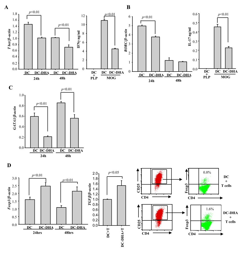
Fig. 3. DC-DHA affects CD4+ T cell differentiation.
DC from C57BL/6 mice were treated with or without 50 µM DHA for 24h, followed by 0.1 µg LPS and pulsing with MOG35–55 for another 24h and extensive washing. DC and DC treated with DHA (DC-DHA) were cultured with MOG35–55-specific CD4+ T cells at 1/20 ratio. 24 and 48h later, T cells were collected, the expression of Tbet (A), RORC (B), GATA-3 (C) and Foxp3 (D, left panel) were assessed by real time RT-PCR. Three days later, supernatants were collected and the production of IFNγ and IL-17 was determined by ELISA (A and B). T cells were collected and subjected to real time RT-PCR to detect expression of TGFβ (D, middle panel). Foxp3+ expression was analyzed by flow cytometry in gated CD4+CD25+ T cells. (D, right panels). One representative experiment of three is shown.

