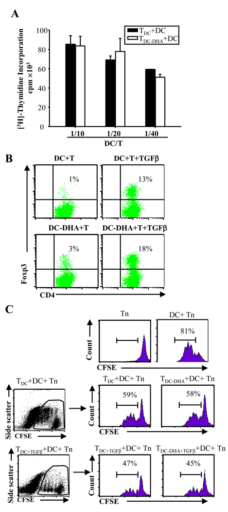
Fig. 4. DC-DHA activated CD4+T cells are not anergic or suppressive.
(A) Activated T cells (TDC or TDC-DHA) were generated from MOG35–55-specific CD4+ T cells cultured with DC or DC treated with docosahexaenoic acid (DC-DHA) pulsed with MOG35–55 and activated with LPS. TDC or TDC-DHA (2×10⁵ cells/well) were cultured with different number of regular DC stimulated with LPS and pulsed with MOG35–55. Three days later, proliferation was measured. (B) Activated T cells were generated from MOG-specific CD4+ T cells cultured with DC or DC-DHA in the presence or absence of 2 ng/ml TGFβ plus 50U IL-2. The Foxp3+ expression was analyzed in the same way as in Fig 3 (D). (C) 1×10⁵ cells/well activated T cells (TDC, TDC-DHA, TDC+TGFβ and TDC-DHA+TGFβ generated from naïve MOG-specific CD4+ T cells cultured with DC, DC-DHA, DC+TGFβ and DC-DHA+TGFβ, respectively) were co-cultured with CFSE-labeled syngeneic MOG-specific naïve CD4+ T cells (Tn) (1×10⁵ cells/well) in the presence of DC (1×10⁴ cells/well) pulsed with 50 µg/ml MOG35–55. T cell proliferation was determined by CFSE dilution using FACS. One representative experiment of three is shown.

