
Figure 6.
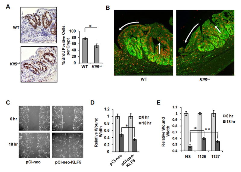
Epithelial proliferation and migration are reduced at sites of ulceration in Klf5+/− mice. Colon tissues were isolated after 5 days of recovery and subjected to immunohistochemical staining. (A) BrdU staining of tissues from mice injected with BrdU 4 hrs prior to sacrifice. Images show crypts in regions adjacent to ulcers. Cell counts are from 6 crypts per mouse, (*P = 0.009; n = 5). (B) Immunofluorescence staining of Ki67 (green) and BrdU (red) in colon tissues from mice injected with BrdU 24 hrs prior to sacrifice. White arrows indicate areas of migration. (C) Wounding assays in DLD-1 cells transfected with empty vector (pCI-neo) or an expression construct for KLF5 (pCIneo-KLF5). (D) Graphical data of relative wound widths from (C). *P = 0.01, n=3. (E) Wounding assays in DLD-1 cells transfected with non-specific siRNA (NS) or two different siRNAs specific for KLF5 (1126 or 1127). *P < 0.001; n=3. **P = 0.03; n=3.