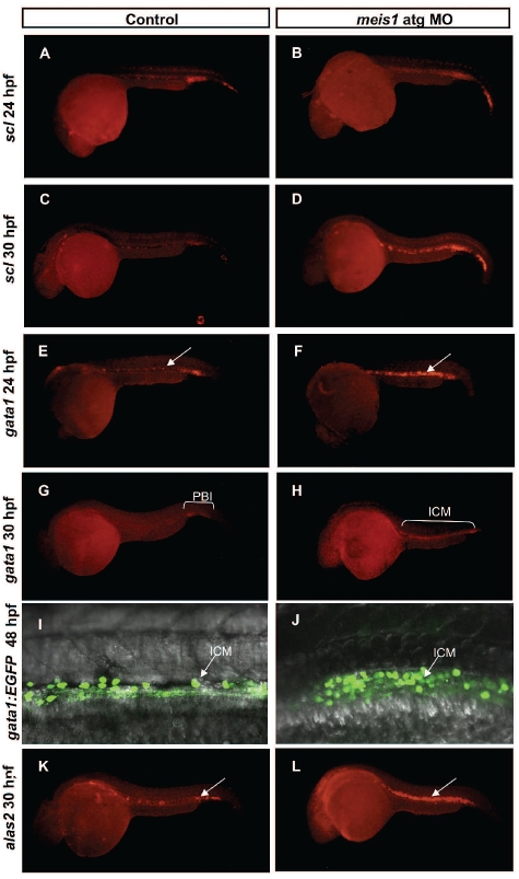
Figure 3.
Meis1 is required for normal differentiation of primitive erythrocytes. (A–B) scl in situ hybridization delineates hematopoietic and endothelial progenitors and the number of scl-positive cells was increased in morphant embryos (B) in comparison with the control (A) at 24 hpf. (C–D) By 30 hpf scl expression in the ICM was 77.5 % increased in MO-injected embryos (D) in contrast to the expression of scl in control siblings (C). To further validate the hematopoietic defect in meis1 MO-injected embryos, expression of erythroid-specific genes was examined. (E–F) In situ hybridization of gata1 at 24 hpf, revealed the 1.5-fold increase of the expression of gata1 in MO-injected embryos (E) in comparison with the control (F). (G–H) However, whereas in control embryos (G) gata1 expression slowly decreased, as gata1-positive cells entered the circulation, in meis1 atg MO-injected embryos (H) it persisted at high levels and was distributed throughout the whole length of the ICM. (I–J) At 48 hpf strong gata1 expression persisted in meis1 MO-injected embryos (J) whereas it was down-regulated in control embryos (I). (K–L) In situ hybridization of the differentiation marker alas2 showed a 3-fold increase in number of alas2-positive cells (L, white arrows point to the ICM, highlighting the accumulation of extra cells in MO-injected embryos) when compared with the controls (K).

