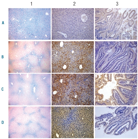
Figure 2.
Cellular localization of iron and BMP6 in the liver and the duodenum of mice with iron overload secondary to an iron-enriched diet or due to Hfe-deficiency. Iron deposits are vizualized by Perls’ staining (1). BMP6 expression was detected by immunohistochemistry in the liver (2) and the duodenum (3). As expected, although Bmp6-deficient mice (A) have the highest iron accumulation, no Bmp6 was detected. C57BL/6 mice with secondary iron overload (B) and DBA/2 Hfe-deficient mice (C) have significant amounts of liver iron and Bmp6 in their liver but not in their duodenum. 129/Sv mice with secondary iron overload (D) have the lowest amount of liver iron and this correlates with Bmp6 staining in the liver. Original magnification x100 (1) or x200 (2 and 3).

