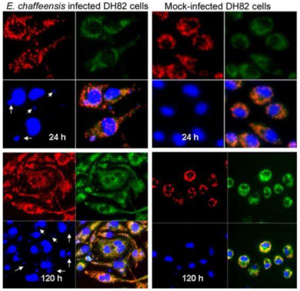
Figure 6.

JC-1 stained E. chaffeensis infected DH82 cells. The images are representatives of E. chaffeensis-infected or mock-infected DH82 at 24 and 120 hours after infection. Red fluorescence shows JC-1 stained mitochondria; green florescence shows JC-1 stained cytoplasm; and blue color is DAPI-stained nuclei and E. chaffeensis morulae (arrows). The image on the bottom-right of each panel is the merged picture. Mock-infected DH82 cells remain as monocytes and became condensed at day 5, but E. chaffeensis-infected DH82 cells became elongated macrophages with multiple nuclei in each cell, suggesting that the infected cells were still multiplying after 5 days of infection in spite of heavy infection with E. chaffeensis. The panels on top are at the same magnification, and the panels on bottom are at the same magnification.
