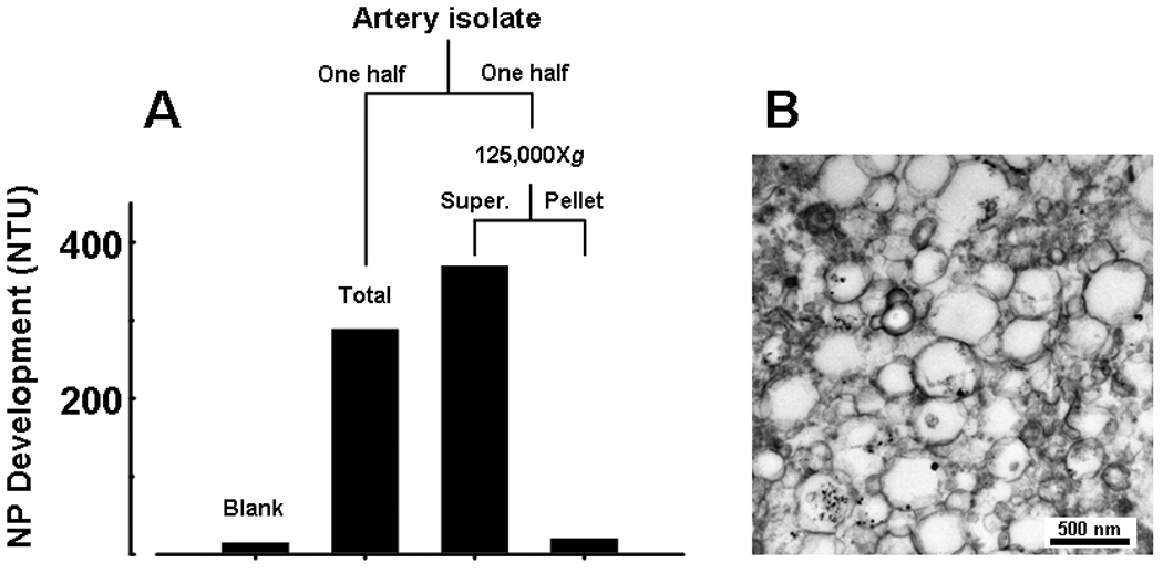
Figure 1.

NPs propagate in vitro from explanted calcific carotid artery isolates after removal of nano-sized components. Fresh artery tissue was prepared as described in Methods, 0.2 µm-filtered and divided in half. One half was added to 5 ml medium and cultured for 3 weeks (Total); the other half was centrifuged at 125,000Xg and the resulting supernate and pellet were cultured separately. A: NP development after 3 weeks in culture: NPs readily propagated from complete sample (Total homog); propagation was similar in replicate sample devoid of structural components (Supernate), but was absent in fraction containing these components (Pellet). B: TEM micrograph showing pelleted structures in size range of matrix vesicles and apoptotic bodies.
