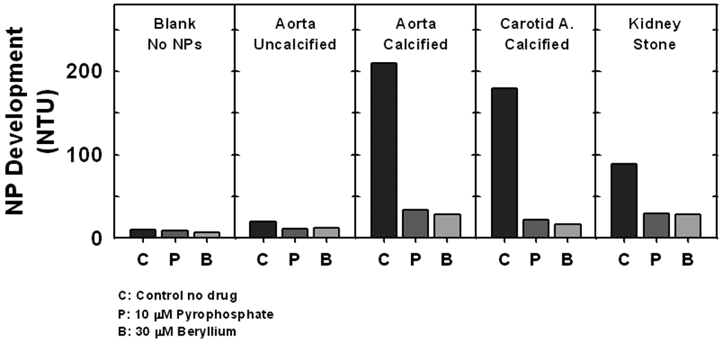
Figure 3.

Propagation of NPs in vitro from isolates of explanted vascular tissue and calcium phosphate kidney stones. Filtered supernatant from the 2,500g spin of tissue homogenates were seeded into flasks. After 14 days in culture, the turbidity of the medium containing total NPs (in Nephelometric Turbidity Units; NTU) was measured and used as an index of NP propagation. NPs propagated from all calcified tissues but not from an uncalcified aorta. Propagation was attenuated by PPi (10 µM), an inhibitor of hydroxyapatite mineral deposition, and by the ALP inhibitor beryllium (30 µM). n=1 each.
