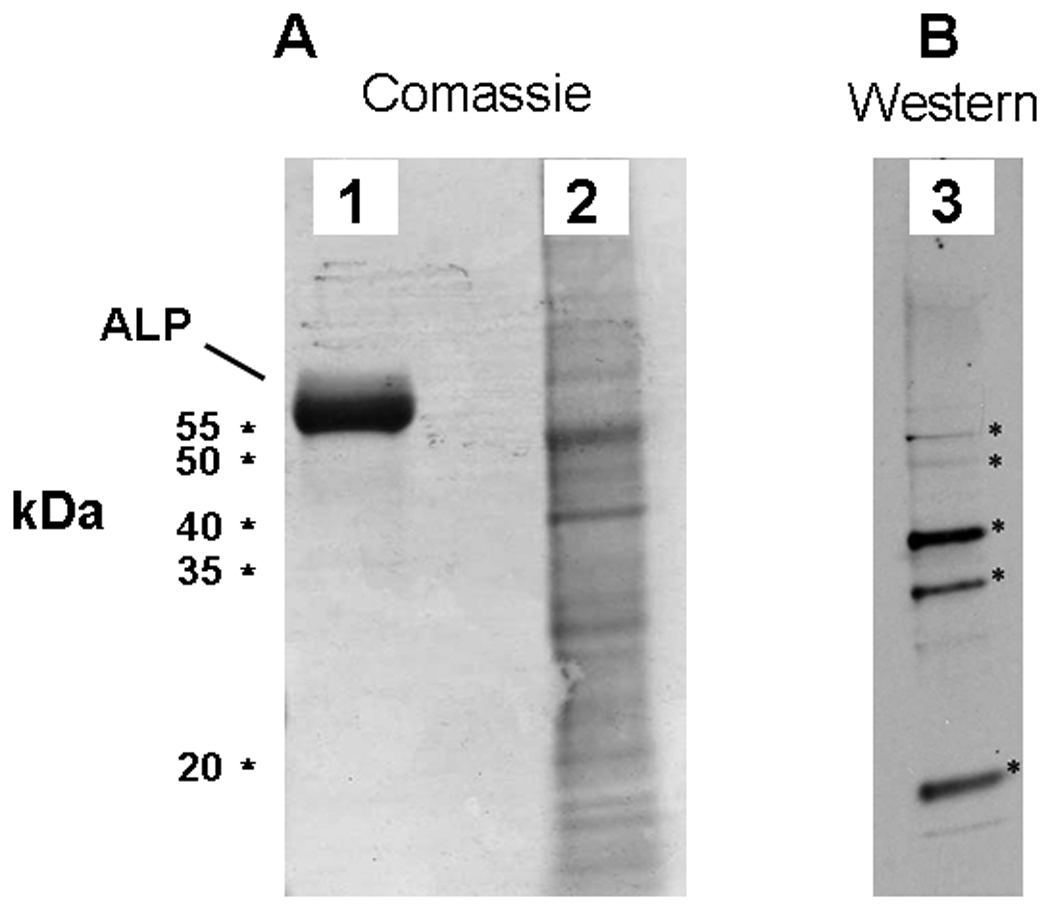
Figure 6.

ALP associates with NPs in vitro. A: Coomassie brilliant blue stained SDS-PAGE gel comparing protein profiles of a decalcified NP isolate (2) and an ALP standard (1). B: Western blot of comparable NP isolate probed with a polyclonal antibody against an internal region of human ALP. Five of the most prominent bands recognized by the antibody (indicated by stars) were cut from a companion gel and subjected to MS/MS analysis for determination of NP protein.
