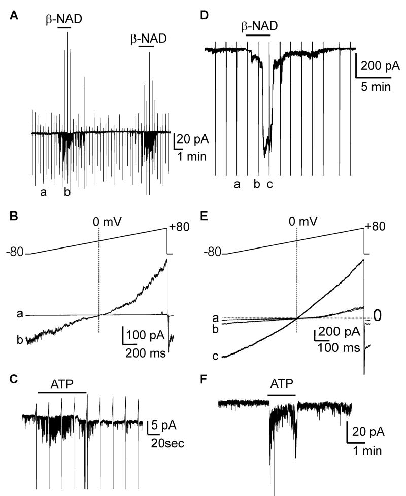
Figure 6. ß-NAD and ATP activate inward currents in SMCs of monkey (A-C) and human (D-F).
(A) In cell-attached patches (pipette CaPSS; bath HK), channel openings were negligible. ß-NAD (1mmol/L) increased openings at -80 mV. (B) Stepping from -80 to +80 mV showed ß-NAD-activated currents reversed at 0 mV in monkey SMC. a (control) and b (ß-NAD) show expanded traces from panel A during ramp depolarization (-80 mV to +80 mV). (C) ATP (1mmol/L) increased channel activity at -80 mV in monkey SMC. (D) Perforated whole-cell conditions (pipette Cs-TEA, bath CaPSS), ß-NAD (5mmol/L) activated inward current at -80 mV. ß-NAD activated-currents reversed upon washout. (E) a, b, and c show expanded traces from panel D during ramp depolarization. ß-NAD activated-currents reversed at 0 mV, demonstrating non-selective cation conductance was activated by ß-NAD. Dotted lines in B and E denote 0 mV and 0 pA. (F) ATP (1 mmol/L) activated inward currents at -80 mV.

