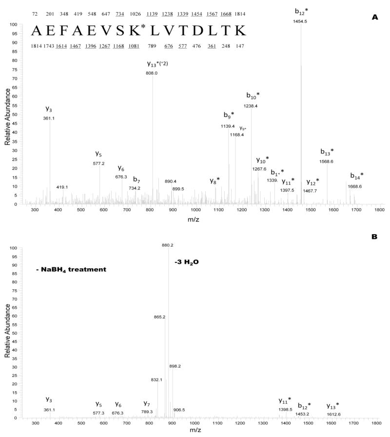
Figure 3.
CID mass spectra recorded on the (M+2H)+2 ion for sodium borohydride pretreated glycated peptide at m/z 908 (A), and for counterpart glycated peptide control not treated with sodium borohydride (B). The sequence of the peptide in (A) is shown with the predicted y and b fragments. Ions observed in spectrum (A) are underlined. K* represents a galactated lysine residue of 292 Da at position 233 in the primary structure of HSA.

