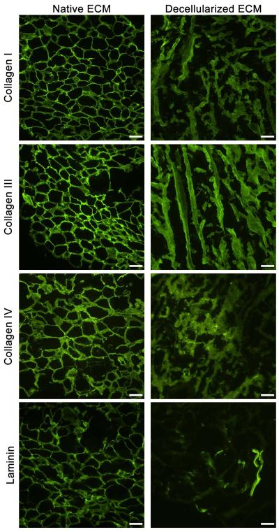
Figure 5. Immunofluorescent Staining of Adipose Matrix.
Fluorescent antibody staining of both fresh human lipoaspirate and adipose matrix decellularized with SDS showed retention of collagens I, III, and IV. Laminin was also present in both cases, but there was some loss of content as a result of the decellularization. Scale bar = 100 μm.

