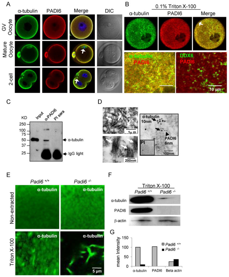
Figure 1. Co-localization and interaction of α-tubulin and PADI6 in murine GV oocytes, mature oocytes, and early embryos.

(A-B) Co-localization of α-tubulin and PADI6 by confocal microscopy before (A) and after (B) treatment with 0.1% of Triton X-100 to release soluble proteins. PADI6 and α-tubulin co-localization is highlighted in merged images. Spindle MTs are indicated by white arrows in panel A. Cortical region is indicated by arrow in panel B. The co-localization of DDX6 and PADI6 serves as a control. (C) Immunoprecipitation of α-tubulin from oocyte extracts using anti-PADI6 antibodies. PI = PADI6 pre-immune sera. (D) Ultra-structure of cytoplasmic lattices (CPLs) and co-localization of α-tubulin and PADI6 at the CPLs by immuno-EM. CPL structures are indicated by white arrows in upper (low magnification) and lower (high magnification) left TEM images and are presented here for reference. Immuno-EM image (right panel) with α-tubulin and PADI6-coated gold particles indicated by black arrows and arrow head, respectively. Inset shows PADI6 pre-immune sera. (E) IIF-confocal analysis of tubulin localization and levels in wild-type and Padi6-null oocytes prior to and following extraction with Triton X-100. (F) Western blot analysis of α-tubulin and PADI6 levels in the insoluble pellet of wild type and Padi6-null oocytes following extraction with Triton. (G) Staining intensity of α-tubulin bands from wild-type and Padi6-null oocytes in Figure F. See also Figure S1, S2 and Table S1.
