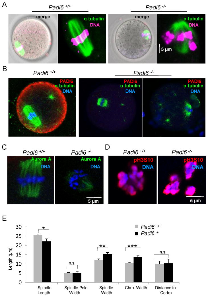
Figure 2. Abnormal meiotic spindle configuration in mature Padi6-null oocytes.

(A) Spindle MT structure in live oocytes. MTs and DNA were stained with Oregon green 488 Taxol and Hoechst 33342 (magenta), respectively. (B) PFA-fixed oocytes were stained with antibodies to α-tubulin (green) and PADI6 (red). (C) Localization of Aurora kinase A (green) on spindles from Padi6 wild-type and mutant oocytes. (D) Same as (C) except oocytes were stained with anti-phospho-histone H3 Ser10 antibody (red). (E) Measurement of spindle apparatus from Padi6 wild-type and mutant oocytes. Chro., chromosome; *, P < 0.05; **, P < 0.001; ***, P < 0.0001; n.s., not significant. Numbers are average ± SEM.
