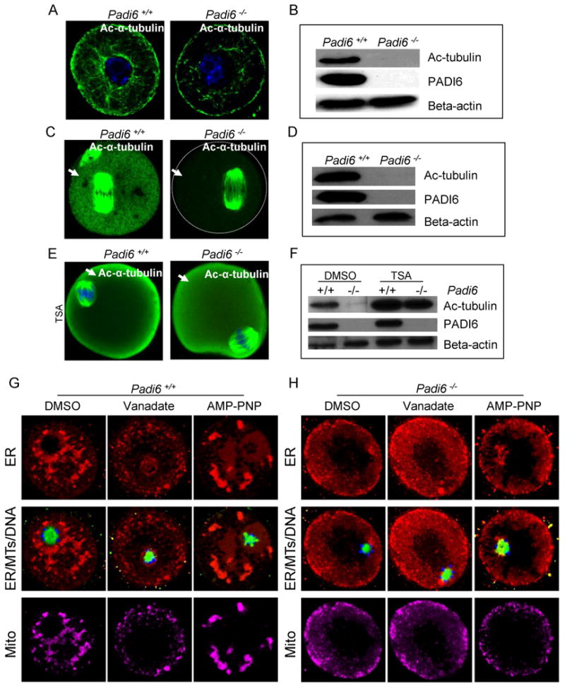
Figure 5. Padi6-null oocytes display reduced levels of cytoplasmic acetylated α-tubulin and treatment of these oocytes with MT motor inhibitors does not affect organelle distribution.

(A-F) Confocal IIF and western blot analysis of acetylated α-tubulin localization and levels in wild-type and Padi6-null GV-stage (A-B), MII-arrested oocytes (C-D), and TSA-treated in vitro matured oocytes (E-F). Cytoplasmic acetylated tubulin was indicated by arrows. (G-H) Effect of MT motor inhibitors on organelle distribution in oocytes. Padi6 wild-type (G) and mutant (H) GV-stage oocytes were cultured with the MT motor inhibitors, vanadate or AMP-PNP, for 3 hours, stained with Tubulin Tracker (green), ER Tracker (red), Mito Tracker (magenta), and Hoechst 33342 (blue), and imaged by confocal microscopy. See also Figure S5.
