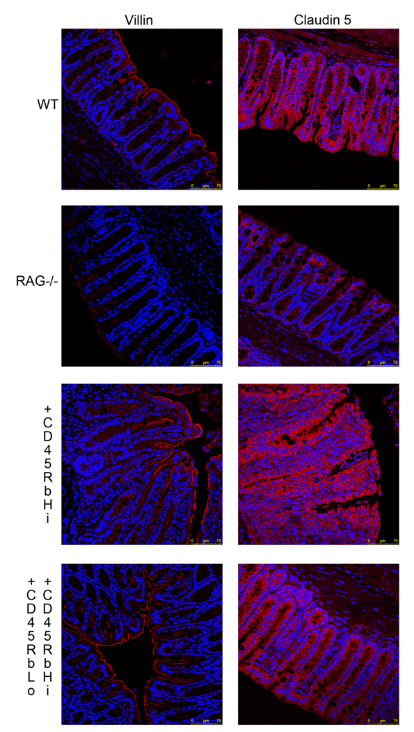
Figure 2.
Colonic tissues sections from C57/BL6 WT, RAG1 deficient mice, as well as RAG1 deficient mice who received either CD4+CD62L+CD45RbHi T cells or CD4+CD62L+CD45RbHi and CD4+CD62L+CD45RbLo T cells were immunostained using an anti-villin (left panel) (red) or anti-claudin 5 (right panel) antibody. The nuclei were stained with Hoechst 33342 (blue). All the samples were analyzed with a Leica SP5-DM Confocal microscope at 40× magnification. These data are representative of 5 experiments.

