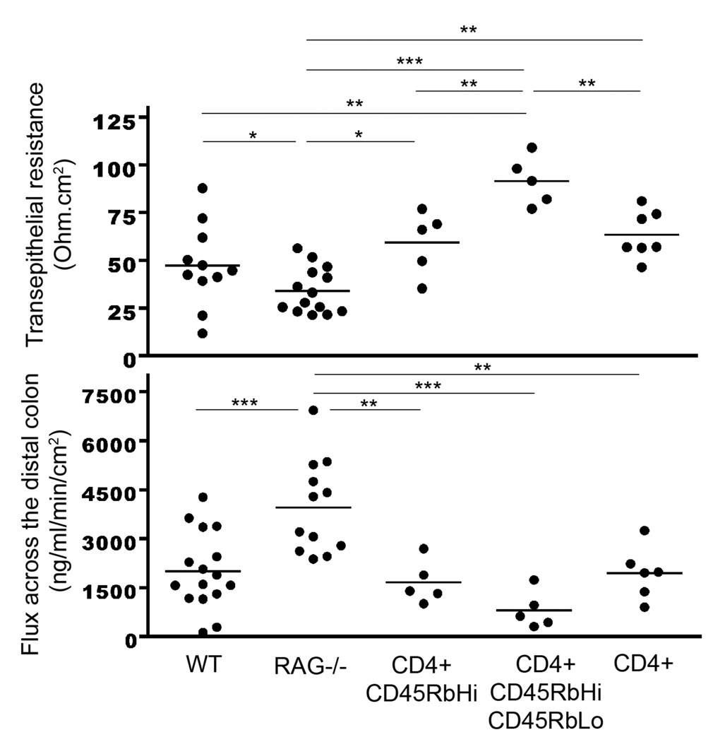
Figure 3.
Assessment of changes in epithelial resistance (top panel) as a measure of passive transcellular and paracellular ion transport, and changes in paracellular permeability (lower panel) for small molecules. WT, non-reconstituted RAG−/− mice, RAG−/− mice reconstituted with CD4+CD62L+CD45RbHi T cells, RAG−/− mice reconstituted with CD4+CD62L+CD45RbHi and CD4+CD62L+CD45RbLo T cells or RAG−/− mice reconstituted with whole CD4+ T cells were sacrificed after 6 weeks. The distal colon was harvested and the resistance was measured along with paracellular permeability (assessed by flux measurements of Dextran-FITC from the mucosal to serosal compartment, measured by spectrofluorometry). Data represent the mean ± SEM of at least 5 mice/group. The symbols *, **, *** refer to statistical significance, respectively <0.05, <0.01, <0.001.

