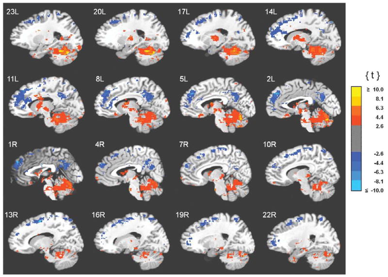
Fig. 2.

Clusters of unique correlations between fMRI activity and 0.05–0.099 Hz EEG spectral power. Functional images are displayed on the standardized average of 27 T1-weighted scans of a single participant known as the "Colin Brain" (Montreal Neurological Institute, Montreal, Canada). Voxel size is 1.0 mm3.
