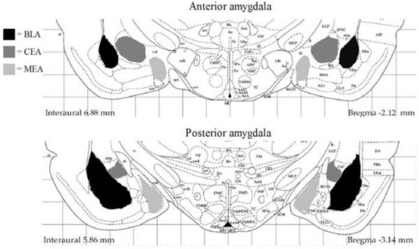
Figure 1.

Depiction of the anatomical levels of the amygdala analyzed in this study. Separate counts were made in the anterior (−1.80 to −2.30 mm from bregma) and posterior (−2.56 to −3.30 mm from bregma) portions of the basolateral/lateral (BLA), central (CEA), and medial (MEA) nuclei of the amygdala. Figure modified from Paxinos and Watson (1998).
