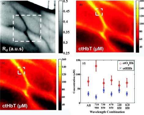
Figure 6.
Reduced spectral content maintains integrity of hemodynamic maps. (a) A single snapshot diffuse reflectance map at 800 nm shows high contrast for veins. (b) SFDI chromophore map produced by a 34-wavelength fit produces comparable maps to (c) two-wavelength fit. The mean and standard deviation of a hemodynamic is calculated over a small ROI covering a vein with multiple wavelength combinations. Wavelength combinations include (1) 34 evenly spaced wavelengths between 650 and 980 nm (all), (2) 710 nm∕980 nm, (3) 730 nm∕850 nm, (4) 670 nm∕850 nm, (5) 670 nm∕850 nm and (6) 730 nm∕850 nm. Combination 1 is fit for four chromophores. Combinations 2–4 are fit for hemodynamic values, with no assumption for water fraction. Underlined combinations 5 and 6 assume a fixed water fraction of 50% to improve fits for hemodynamic parameters. A 670 nm∕850 nm pair with assumed water and lipid fractions performs the best.

