
Figure 2. FoxO1 binds the promoter region of Sestrin3 gene and directly induces its mRNA levels.
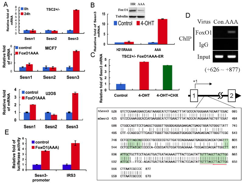
A. FoxO1 increases Sesn3 mRNA level. Total RNA was isolated from Tsc2+/- MEFs stably expressing FoxO1(AAA)-ER in the absence or presence of 4-OHT or from FoxO1(AAA) adenovirus or control adenovirus infected MCF7 and U2OS cells. mRNA levels were quantified by qRT-PCR as described in Experimental Procedures. Fold change in mRNA levels was calculated by normalizing to actin mRNA. B. DNA binding deficient FoxO1 cannot induce Sesn3 mRNA. Total RNAs were extracted from Tsc2+/-cells stably expressing FoxO1(AAA)-ER or FoxO1H215R(AAA)-ER in the absence or presence of 4-OHT. Relative mRNA levels were determined by qRT-PCR. Insert shows the levels of FoxO1AAA and FoxO1H215R(AAA). C. The induction of Sesn3 mRNA by FoxO1 does not require de novo protein synthesis. Tsc2+/- FoxO1(AAA)-ER cells were treated with cycloheximide (CHX) for one hour prior to the addition of 4-OHT to prevent de-novo protein synthesis. After addition of 4-OHT for four hours, relative levels of mRNA were determined by qRT-PCR. D. FoxO1 directly binds to the promoter region of the Sesn3 gene. U2OS cells were infected with FoxO1(AAA) adenovirus and then subjected to a ChIP assay using FoxO1 antibodies, as described in Experimental Procedures. The localization of the binding region within the first intron of Sesn3 gene is indicated schematically. Sequences show four predicted consensus FoxO binding sites highlighted within the binding region. The high homology of these sequences between the mouse and human Sesn3 genes is shown. E. FoxO1 regulates transcription from the Sesn3 promoter in a luciferase reporter assay. FoxO1(AAA) MEFs were transfected with pGL3-Sesn3 promoter or conventional pGL3-IRS3 (three copies of insulin response element of IGFBP1 promoter) construct as a positive control. Cells were treated with or without 4-OHT and subjected to Dual-Luciferase reporter assay (Promega). Luciferase activities were normalized to a co-expressed Renilla luminescent signal, and is shown as relative folds against control samples.