
Fig. 5.
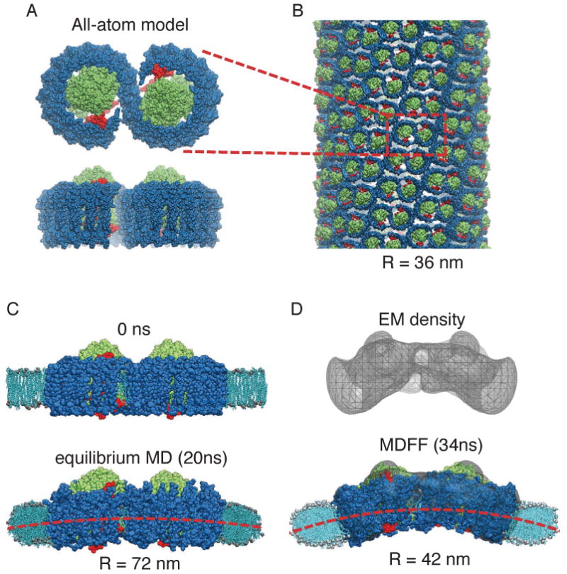
Membrane-bending properties of the bacterial photosynthetic core complex. (A) Top (perpendicular to the membrane plane) and side (along the membrane plane) views of an all-atom model of the core complex dimer [36] (RC: green; LH1: blue; PufX: red). (B) Example of a tubular photosynthetic membrane observed in mutant Rba. sphaeroides [44]. The core complex dimers can be seen to arrange helically in the tubular vesicle, and one complex is outlined in red for identification. This tubular membrane has a radius of 36 nm. (C) Setup of the membrane-protein system for equilibrium MD of the all-atom model. The membrane is shown in light blue; the water box included in the simulation is not shown for clarity. During an equilibrium MD simulation, a small bending was seen in the core complex dimer and a shallow curvature in the membrane emerged [36]. (D) 25Å-resolution EM reconstruction obtained through single-particle analysis [44]. The all-atom model was fitted into the EM map employing MDFF; during the MDFF simulation the surrounding membrane developed a more prominent curvature [11] that remained stable in an equilibrium simulation.