
Figure 3.
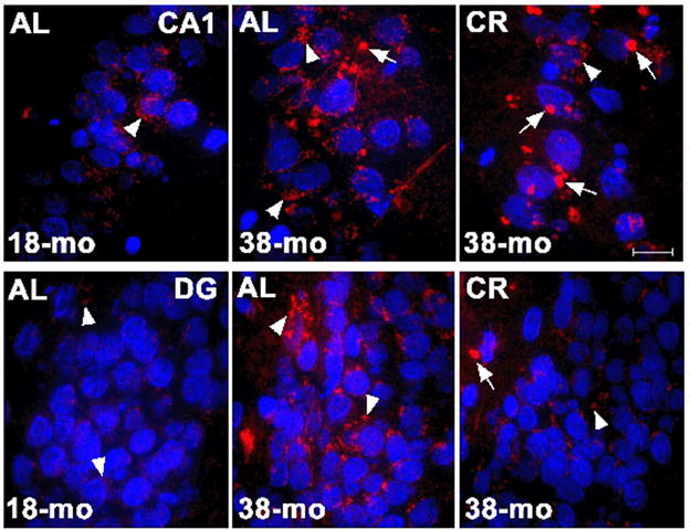
Immunofluorescence showing ubiquitinated inclusions (red) in CA1 (top row, 60X) and DG (bottom row, 60X) regions for 18 mo AL (left column), 38 mo AL (center column) and 38 mo CR (right column). Nuclei were stained with a Hoechst (blue). There appears to be more ubiquitin staining in 38 AL compared to 18 AL and the ubiquitin was diffuse (arrow heads) and distributed throughout the cell body. In comparison, overall ubiquitin-like immunoreactivity appears reduced by CR and more focal, often limited to inclusions (arrows) adjacent to nuclei. The calibration bar is 10 μM.