
Table 1.
Structural analysis of P450cam conformers.
| Substratea | PDB | PC1b | PC2b | F helix shiftc (Å, deg) | G helix shiftc (Å, deg) | FG helix angle (deg) | Axial Water | Missing B′ helix | Ref |
|---|---|---|---|---|---|---|---|---|---|
| P450CAM-C | |||||||||
| Camphor |
2CPP | 25.7 | 2.9 | — | — | −148.9 | No | — | (4) |
| 1YRC | 29.4 | 4.9 | 0.28, 2.7 | 0.25, 2.8 | −149.4 | (42) | |||
| 5CP4 | 24.8 | 3.7 | 0.22, 2.7 | 0.40, 1.8 | −150.1 | (43) | |||
| 2ZAX | 28.7 | 5.7 | 0.23, 3.1 | 0.28, 2.5 | −148.7 | (44) | |||
| 1DZ4 | 26.7 | 6.5 | 0.11, 1.2 | 0.55, 0.9 | −150.1 | (45) | |||
| 1S-camphor |
1AKD | 23.9 | 6.1 | 0.41, 2.7 | 0.48, 1.4 | −149.0 | No | — | (46) |
| Imidazole |
2H7Q | 24.1 | 4.9 | 0.26, 2.2 | 0.54, 1.5 | −151.4 | No | — | (47) |
Nicotine
|
1P2Y | 25.9 | 5.6 | 0.08, 5.2 | 0.36, 0.5 | −148.2 | No | — | (48) |
Metyrapone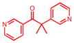
|
1PHG | 25.6 | 3.2 | 0.07, 1.4 | 0.12, 0.5 | −148.6 | No | — | (49) |
| P450CAM-I | |||||||||
| AdaC1-C8-Dans |
1RE9 | 6.4 | −9.9 | 3.30, 17.8 | 1.00, 5.9 | −156.6 | No | — | (66) |
| 1LWL | 4.4 | −11.5 | 3.59, 17.4 | 1.33, 5.4 | −156.5 | (32) | |||
| 3P6M | 2.7 | −11.2 | 3.77, 15.5 | 1.52, 5.4 | −156.7 | This study | |||
| 3P6N | 2.2 | −11.1 | 3.75, 16.0 | 1.50, 6.2 | −157.2 | This study | |||
| AdaC1-C8EtgGlu-Bio |
3OIA | 3.0 | −11.6 | 3.71, 15.8 | 1.27, 7.3 | −153.2 | No | — | Unpublished |
| AdaC1-Etg-Dans |
3P6O | 3.7 | −10.6 | 3.73, 16.7 | 1.42, 6.0 | −153.5 | No | — | This study |
| AdaC1-C6-Bio |
3P6P | 2.5 | −10.3 | 3.91, 15.2 | 1.37, 6.6 | −157.8 | No | — | This study |
| 3OH-AdaC1-C8-Dans |
3OL5 | 1.1 | −10.5 | 3.88, 14.9 | 1.96, 5.1 | −155.8 | Yes | — | Unpublished |
| P450CAM-O | |||||||||
| AdaC1-C4-Dans |
1RF9 | −9.9 | 0.7 | 3.94, 15.9 | 3.68, 10.8 | −149.7 | Yes | — | (66) |
| AdaC2-Etg-Boc |
3P6Q | −15.6 | 3.2 | 4.51, 17.2 | 3.95, 15.2 | — | Yes | 92–95 | This study |
3OH-AdaC1-Etg-Boc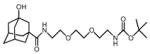
|
3P6R | −16.3 | 4.1 | 4.84, 16.6 | 4.57, 15.2 | −151.1 | No | 90–96 | This study |
| AdaC2-C8-Dansd |
3P6S | −17.3 | 2.3 | 4.59, 17.1 | 4.46, 14.9 | −150.2 | No | — | This study |
| 3P6T | −15.5 | 0.7 | 4.24, 16.8 | 4.28, 15.7 | −150.6 | This study | |||
| AdaC3-C6-Dans |
3P6U | −21.0 | 3.8 | 4.95, 17.8 | 4.83, 17.4 | −150.6 | Yes | 93–94 | This study |
3Et-AdaC1-Etg-Boc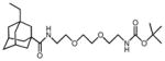
|
3P6V | −20.4 | 4.6 | 5.06, 17.4 | 5.02, 16.0 | −150.7 | No | 90–96 | This study |
| AdaC3-Etg-Boc |
3P6W | −20.5 | 3.8 | 5.04, 17.1 | 4.85, 17.4 | −151.0 | No | 91–95 | This study |
| AdaC3-C8-Dans |
3P6X | −22.2 | 3.9 | 5.07, 18.7 | 4.86, 18.1 | −150.4 | Yes | 90–94 | This study |
AdaC1-perfluorobiphenyl-Ru(bipy)3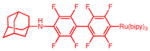
|
1K2O | −22.7 | 3.8 | 5.54, 21.0 | 5.14, 18.8 | −151.6 | Yes | — | (65) |
| AdaC1-C8-Ru(bipy)3 |
1QMQ | −16.0 | 3.3 | 4.43, 16.0 | 4.28, 14.7 | −149.0 | Yes | — | (64) |
| Substrate-free | 3L61 | −24.9 | 4.3 | 5.27, 19.3 | 5.09, 17.9 | −150.2 | Yes | 90–97 | (18) |
| 3L62 | −21.5 | 3.6 | 4.93, 18.2 | 4.73, 16.9 | −150.2 | 91–94 | (18) | ||
a
Well ordered electron density included for portions of the model shown in red.
b
PC1 and PC2 are first two principal components in the PCA of crystal structures described in this table.
c
Helix shift is calculated relative to the camphor-bound closed conformation (PDB entry 2CPP) at the center of the F helix (Lys178) and G helix (Lys197).
d
The entire substrate was only resolved in the structure of PDB entry 3P6T.