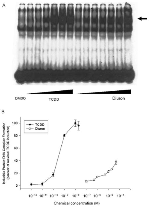
FIGURE 5.
Dose-dependent stimulation of AhR transformation and DNA binding by TCDD and diuron in vitro. Guinea pig hepatic cytosol (8 mg protein/mL) was incubated with DMSO (20 μL/mL, final concentration) or increasing concentrations of TCDD (1, 10, 100 pM and 1, 10, 20 nM) or diuron (0.1, 0.5, 1, 5, 10, 20, 50 μM) for 2 h at 20°C. Protein–DNA complexes were resolved by gel retardation analysis (a) and the amount of induced protein–DNA complex formation determined by phosphorimager analysis (b). Values are expressed in the figure as the percentage of maximal TCDD induction and represent the mean ± SD of triplicate determinations. Induced complex formation at all concentrations of TCDD ≥10−11 M and of diuron ≥10−7 M were significantly greater than the DMSO-treated sample at p < 0.01 as determined by Student’s t-test. The arrow indicates the position of the AhR:DRE complex.

