
Figure 5.
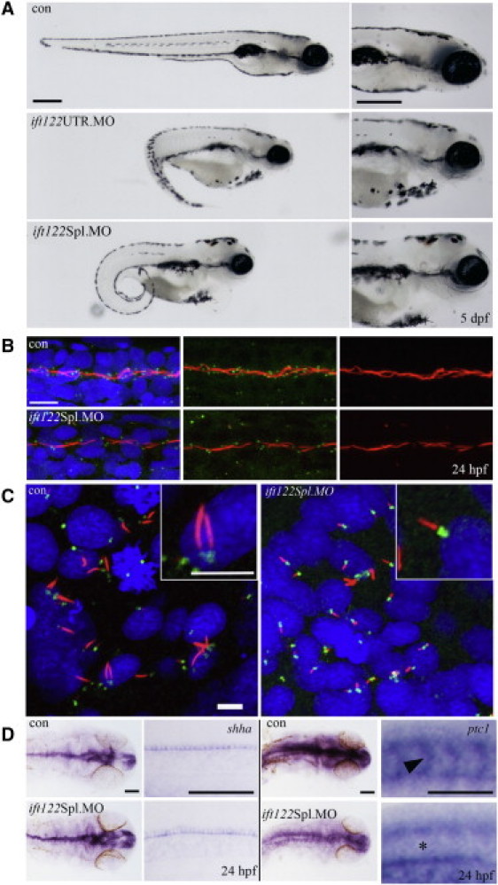
Loss of ift122 in Zebrafish Results in a Ciliopathy-Related Phenotype
(A) Gene-specific knockdown by morpholino modified antisense oligonucleotides to either the 5′UTR or to the exon 1-intron 1 splice boundary of ift122 caused reduced ocular development, melanocyte mislocalization, curly tails, renal cysts, and heart edemas by 5 dpf. All common features related to loss of ciliary genes in zebrafish. The scale bar represents 400 μm.
(B) Ift122 morphants display reduced and shortened motile cilia. Immunofluorescence staining for cilia (acetylated α-tubulin, red), centrosomes (γ-tubulin, green), and nuclei (DAPI, blue) in the pronephric duct (PND) shows reduced centrosome numbers and interrupted and/or shortened cilia in morphant compared to control embryos. The scale bar represents 10 μm.
(C) Cilia in the KV of ift122 Spl. Morphants are visibly shorter than uninjected controls. The scale bar represents 5 μm.
(D) A mild reduction in Hh signaling was observed in morphant embryos. Embryos were analyzed for either shha or ptc1 by in situ hybridization at 24 hpf; images of the head and somites (at approximately the somite 15 position) are displayed for comparison. Shha accumulates in the floor plate of the neural tube and in the notochord (at low levels) at the 24 hpf stage; this expression remains unperturbed in morphant embryos. Ptc1 appears to be globally reduced in ift122 morphants, most notably in the somites (see asterisk) where ptc1 is normally abundant in uninjected control embryos (see arrowhead). The scale bar represents 100 μm.